Download attachment:PDF
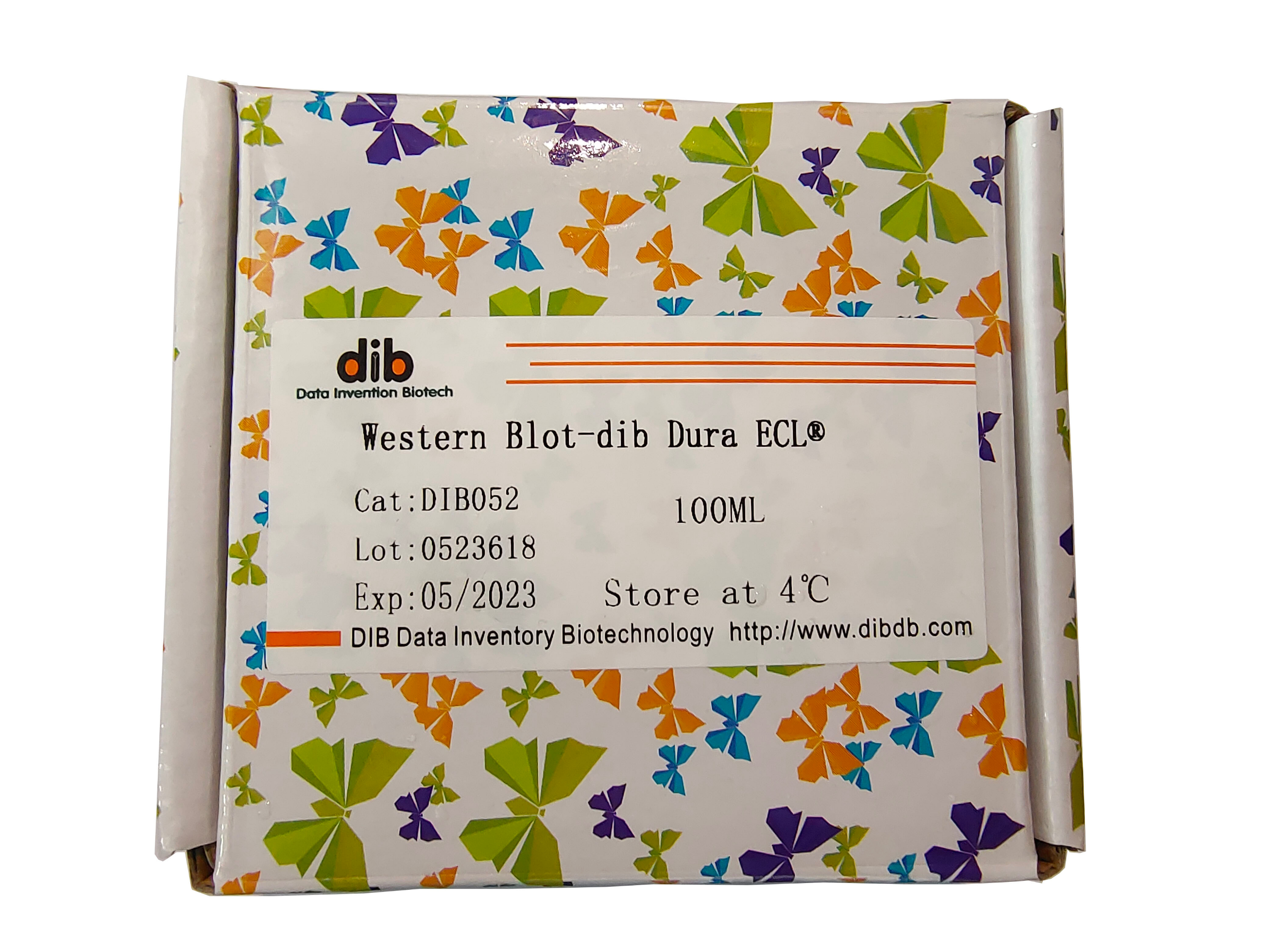
Dura ECL Super Sensitive Luminescent Liquid Manual
Product number:DIB052-100ml DIB052-500ml
Product specifications:100ml 500ml
Storage: Transport at room temperature, store reagents at 4°C and protected from light after receipt
Dura ECL is an enhanced chemiluminescence (ECL) substrate with ultra-high sensitivity and an ECL luminescent solution with ultra-long luminescence time. It is also currently the most sensitive chemiluminescence substrate of Fubaike, with a luminescence time of up to 16h.Horseradish peroxidase (HRP) can be used to achieve low femtogram level western blot detection.The luminescent substrate system designed based on the "ping-pong" mechanism is adopted due to the unique core patented technology of Fu Baike.HRP can realize low femtogram level western blot detection, and its sensitivity, luminescence time and background are significantly better than T company and M company.
Product introduction ura ECL's substrate with the highest sensitivity and longest luminescence time is an enhanced chemiluminescence substrate for horseradish peroxidase (HRP), which has the following characteristics:
• Sensitive—use appropriate primary antibodies and In the case of the secondary antibody, the protein bands with femtogram abundance can be detected on the nitrocellulose membrane or PVDF membrane
• Quantification-the quantitative detection range of the obtained signal spans two orders of magnitude
• Bright signal-exposure through film or imaging system, Easy to capture images
• Long signal duration-under optimized conditions, the detectable optical signal output can be up to 16 hours
• Stable reagents-kit components can be stored stably at 4°C for one year, and stable at room temperature 6 Months
• Economical price-the formulation is optimized for the detection of very low concentrations of antibodies
• 1ng to 0.2 µg/mL primary antibody (diluted 1:5,000 to 1:100,000 times with 1 µg/mL stock solution)
• 2ng to 10 ng/mL secondary antibody (diluted 1:100,000 to 1:500,000 times with 1 µg/mL stock solution)
When Dura ECL substrate is used with optimized antibody concentration and blocking buffer, low-abundance target proteins that cannot be detected by conventional ECL substrates can be detected.
Important note:
Ø For best results, all components of the system must be optimized, including sample volume, primary and secondary antibody concentrations, and types of membranes and blocking reagents.
Ø The antibody concentration required for the detection of this product is lower than that of the precipitation colorimetric HRP substrate.To optimize antibody concentration, perform a systematic dot blot analysis.
Ø No one blocking reagent is optimal for all systems, so it is very necessary to find the most suitable blocking buffer for each western blot detection system.The blocking reagent may cross-react with the antibody, resulting in non-specific signals.The blocking buffer also affects the sensitivity of the system.When switching from one substrate to another, signal attenuation or background increase sometimes occurs. The reason may be that the blocking buffer is not suitable for the new detection system.
Ø When using avidin/biotin detection system, avoid using milk as a blocking reagent, because milk contains unquantified endogenous biotin, which will cause high background signals.
Ø Ensure the use volume of washing buffer, blocking buffer, antibody solution and substrate working solution to ensure that the blotting membrane is completely covered by the liquid during the entire experiment and to prevent the membrane from drying out.Increasing the amount of blocking buffer and washing buffer used can reduce non-specific signals. For best results, use a shaker during the incubation step.
Ø Add Tween20 (final concentration 0.05-0.1%) to blocking buffer and diluted antibody solution to reduce non-specific signals.Use high-quality products such as detergents.It is kept in ampoules, and the content of peroxides and other impurities is very low.
Ø Do not use sodium azide as a preservative in the buffer.Sodium azide is an inhibitor of HRP. Avoid direct contact between hands and the membrane. Wear gloves or use clean tweezers during the experiment. All equipment must be clean and free from foreign substances.Metal instruments (such as scissors) must not have visible rust.Rust may cause spot formation and high background.
Ø The substrate working solution can be stable for 8 hours at room temperature.Sunlight or any other strong light may damage the substrate. For best results, store the substrate working solution in an amber bottle and avoid long-term exposure to any strong light and short-term exposure to the laboratory's routine lighting Will not damage the working fluid
Other required materials
l The blotting membrane that has been transferred: Separate the proteins with a suitable electrophoresis method and transfer these proteins to the nitrocellulose membrane.l Dilution buffer: use Tris or phosphate buffer.
l Washing buffer: add 5mL 10% Tween-20 to 1000mL dilution buffer (the final concentration of Tween-20 will be 0.05%).
l Blocking reagent: add 0.5mL of 10% Tween-20 to 100mL of blocking buffer, and select a blocking buffer with the same basic components as the dilution buffer.
l Primary antibody: Choose an antibody specific to the target protein.Use dilution buffer to prepare a stock solution of the antibody.Use blocking reagent to dilute the antibody from the stock solution to the antibody working solution.The optimal dilution depends on the primary antibody and the amount of antigen on the membrane.
l HRP-labeled secondary antibody: Choose a HRP-labeled secondary antibody that specifically binds to the secondary antibody, and use the dilution buffer to prepare the antibody storage solution.Use blocking reagent to dilute the antibody from the stock solution to the antibody working solution.The dilution is between 1:100,000 and 1:500,000 or the concentration of the antibody working solution is 2-10ng/ml.This concentration range also applies when using streptavidin-HRP.The optimal dilution of the secondary antibody depends on the HRP-labeled secondary antibody and the amount of antigen on the membrane.
l Film cassette, developing and fixing reagents for processing radiographic films l Rotary shaker for incubation.
Detailed operation steps of western blotting method
1) Remove the imprinting membrane from the protein transfer equipment, add a suitable blocking solution and incubate in the greenhouse for 20-60 minutes while shaking.To block non-specific protein binding sites on the membrane.Please note: It is very important to use the antibody dilution recommended in the previous section.
2) Remove the membrane from the blocking solution and incubate it with the working solution of the primary antibody in the greenhouse for 1 hour while shaking; or incubate overnight at 28°C without shaking.
3) Add sufficient washing buffer to the membrane to ensure that the buffer covers the membrane completely.Incubate with shaking for ≥5 minutes, change the washing buffer and repeat this step 4-6 times.Increasing the volume of the wash buffer, the number of washes and the washing time help to reduce the background signal.Note: Before incubation, a short rinsing of the membrane in the washing buffer will improve the washing efficiency.Please note: It is very important to use the HRP-labeled secondary antibody dilution suggested above.
4) Incubate the HRP-labeled secondary antibody working solution with the membrane in the greenhouse for 1 hour while shaking.
5) Repeat step 3 to remove unbound HRP-labeled secondary antibody.Note: The membrane must be washed thoroughly after incubating with the HRP-labeled secondary antibody.
6) Mix A solution and B solution in equal proportions to prepare a working solution.Use 0.01~0.1ml working solution per cm2 of membrane.The working fluid can be stable for 8 hours in the greenhouse.Note: Heavy rain, sunlight or any other strong light may damage the working fluid. For best results, store this working fluid in an amber bottle and avoid any long-term heavy rain.Common lighting in the laboratory will not harm the working fluid.
7) Incubate the imprinted membrane in the working solution for 5 minutes.
8) Take out the imprint film from the working fluid and place it in a plastic sheet or clean plastic paper (film). Use a piece of absorbent paper to absorb the excess liquid, and carefully press out bubbles from the imprint and the plastic paper .
9) Place the imprint film wrapped in plastic paper (film) in a film cassette with the protein side facing up, and turn off all lights except for the lights suitable for film exposure (such as red safety lights).
Note: The film must be kept dry during exposure. For best results, take the following measures: * Ensure that the excess substrate is completely removed from the film and plastic paper.* During the entire film processing, use gloves.* Do not place the imprint film on the developed film, because the chemicals on the film will weaken the signal.
10) Place the X-ray film on top of the film.It is recommended that the first exposure is 60 seconds.The exposure time can be adjusted later to achieve the best results.The chemiluminescence reaction is strongest during the first 5-30 minutes after the substrate incubation.This reaction can last for several hours, but the intensity will decrease with time. If the substrate is exposed for a longer time after incubation, the exposure time may need to be extended to obtain a stronger signal.If you use phosphorescent storage imaging devices (such as Bio-Rad's molecular imager system) or CCD cameras, longer exposure times may be required.Warning: Any movement between film and film may cause artificial, non-specific signals on the film.
11) Use suitable developer and fixer to develop the film.If the signal is too strong, shorten the exposure time or peel off the imprinting film and reduce the antibody concentration to retest.
Common problems and solutions
| problem |
Possible problem |
solution |
| Reversal image on the film (ie black background, white band) Brown or yellow band mark on the film The luminous signal duration in the dark room is less than 8 hours |
Too much HRP in the system |
Dilute the HRP-labeled secondary antibody at least 10 times |
| Weak or no signal |
Too much HRP in the system depletes the substrate and causes the signal to decay rapidly |
Dilute the HRP-labeled secondary antibody at least 10 times |
| Insufficient amount of antigen or antibody |
Increase the amount of antibodies or antigens |
| Low protein transfer rate |
Optimize transfer |
| Low HRP or substrate activity |
See note below |
| High background |
Too much HRP in the system |
Dilute the HRP-labeled secondary antibody at least 10 times |
| Insufficient closure |
Optimize sealing conditions |
| Enclosed machine is not suitable |
Try a different blocking reagent |
| Insufficient washing |
Increase washing time, number of times or washing buffer volume |
| Film overexposed |
Reduce exposure time or use background remover |
| The concentration of antigen or antibody is too high |
Reduce the amount of antibodies or antigens |
| Spots in protein bars |
Low protein transfer efficiency |
Optimize the transfer process |
| Uneven hydration of the membrane |
Properly hydrate the membrane according to the manufacturer’s recommendations |
| There are bubbles between the film and the film |
Before film exposure, remove air bubbles |
| The film has spots on the background |
There are aggregates in the HRP-labeled secondary antibody |
Use 0.2um filter |
| Non-specific band |
Too much HRP in the system |
Dilute the HRP-labeled secondary antibody at least 10 times. |
| Non-specific protein binding caused by SDS |
SDS is not used during testing |
*To detect the activity of the system, prepare 1-2ml of working solution of the substrate in a clean test tube in a dark room.Turn off the light and add 1ul undiluted HRP-labeled secondary antibody working solution.The solution should emit blue light immediately, and the blue light signal fades in the next few minutes.
| product name |
Detection concentration |
Recommended antibody incubation concentration(ng/mL) |
| ECL plus |
Pick grade~10-12g |
Primary antibody concentration:100~500
Concentration of secondary antibody:10~50 |
|
| West Pico ECL |
Less than picogram level <10-12g |
Primary antibody concentration:100~500
Concentration of secondary antibody:10~50 |
|
| West Femto ECL |
Less than femtogram<10-15g |
Primary antibody concentration:20~100
Concentration of secondary antibody:4~20 |
|
| West Dura ECL |
Less than femtogram<10-15g |
Primary antibody concentration:2~100
Concentration of secondary antibody:2~10 |
|
Product parameter
| Name |
Pico ECL |
| CAT# |
DIB052-100mL;DIB052-500mL |
CAS# |
N/A |
| Storage# |
4°C dry and avoid light |
Shelf Life# |
12 months |
| Ex(nm)# |
N/A |
Em(nm)# |
N/A |
| MW# |
N/A |
Solvent# |
N/A
|

